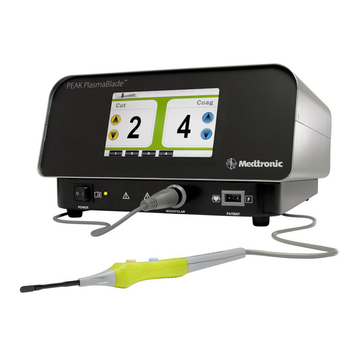
Plasma generator Pulsar II MEDTRONIC
August 2, 2019
Neurosurgical drill Midas Rex Medtronic
August 2, 2019Intracranial neuroendoscope Lotta KARL STORZ
Σύστημα Ενδοσκοπίου LOTTA SYSTEM του οίκου KARL STORZ για επεμβάσεις εγκεφάλου (Εξεργασία Ενδοκοιλιακού Όγκου και ενδοσκοπικής ατραυματική διαστολή της 3ης κοιλίας
The LOTTA® system has been designed to perform the full range of endoscopic intracranial interventions in adults and children. The cornerstone of the system is based on the two ventriculoscopes Little LOTTA® and LOTTA®. These enable the treatment of all forms of obstructive hydrocephalus, intraventricular tumors and cysts as well as arachnoid and intraparenchymal cysts. An all-round solution, the LOTTA® system offers a free choice between the Little LOTTA® with its smaller diameter, more convenient handling and use in a wide range of applications such as ventriculostomies, septostomies, tumor biopsies and cyst fenestrations and the LOTTA® with its larger dimensions, which is not only suitable for the therapies mentioned above but is also particularly effective for the removal of colloid cysts, tumor resections, stent implantations as well as aqueductoplasties with subsequent stenting.
The somewhat larger diameter of the LOTTA® ventriculoscope allows the surgeon to perform bimanual dissection using two instruments. These can be used simultaneously
in separate channels to enable more technically sophisticated procedures. Furthermore, the resection of larger tissue samples is possible, which benefits therapies such as tumor resection or cyst removal.
All intracranial procedures can thus be carried out. However, there are situations where
a 30° viewing angle proves useful. A 30° viewing angle directed on the working channel allows earlier visualization of instruments. Therefore, the use of the LOTTA® 30° in narrow structures is beneficial. In addition, neighboring structures can easily be viewed during resections of cysts or tumors, for example, during the treatment of colloid cyst of the attachment point at the tela choroidea in the roof of the 3rd ventricle.
he ventriculoscopes are equipped with a HOPKINS® wide-angle straight forward telescope with a high light-transmitting capacity which delivers unsurpassed image quality and safe orientation, even in protein-rich or bloody CSF fluid. The central working channel is flanked on both sides with two side channels with a smaller diameter. One is used for irrigation/suction and the other for the use of a second instrument.
The irrigation function ensures that continuous cleaning is maintained in the area in front of the endoscope, even when visibility is hindered (cloudy CSF in the case of ventriculitis and/or ventricle bleeding). The drainage channel always remains open to prevent critical intracranial pressure increase caused by excessive irrigation. To facilitate insertion of the instruments into the working channel, a funnel-shaped enlargement has been integrated at the entrance to the working channel. Thanks to this stable construction, both ventriculoscopes are less susceptible to damage during cleaning, sterilization and storage.


