
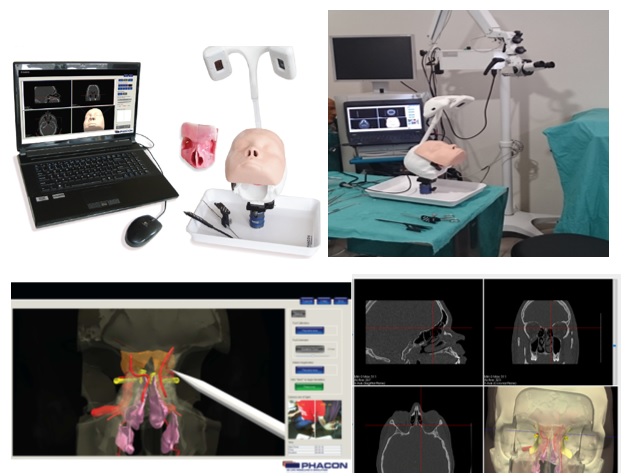
Training in anatomical preparations under intraoperative navigator guidance with CT scan in 3 sections and visualization of the 3D model with the possibility of normal or endoscopic viewing mode, with or without an instructor.
Key Features
-
- Using educational material before and during training.
- The model is suitable for intraoperatively guided Fess, optic nerve decompression, transsphenoidal approach for hypophysectomy and orbital decompression.
- It features bone with features that simulate real conditions, ethmoid alveoli, sphenoid, sinuses and frontal sinuses, faithful simulation of the nasal mucosa, flexible turbinates, optic nerve, pituitary gland, carotid and sphenoid arteries, ethmoid hook, ethmoid osteocyst, etc.
- Available in various variations: with normal anatomy, anatomy with polyps, anatomy with shallow skull base, anatomy for septoplasty and rhinoplasty.
- Critical anatomical structures can be highlighted with different colors on the navigator screen so that they are distinguishable during surgical access.
- The phacon trainer system software features an automatic recorder and audible signaling of the damage that will be caused to critical anatomical structures during each user's surgical access, with the ability to store and compare them with subsequent training.